Acute Pain and Anxiety related to Pyelonephritis

Acute Pain and Anxiety related to Pyelonephritis

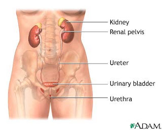
Pyelonephritis is a type of urinary tract infection (UTI) that affects one or both kidneys.

Pyelonephritis is caused by a bacterium or virus infecting the kidneys. Though many bacteria and viruses can cause pyelonephritis, the bacterium Escherichia coli is often the cause. Bacteria and viruses can move to the kidneys from the bladder or can be carried through the bloodstream from other parts of the body. A UTI in the bladder that does not move to the kidneys is called cystitis.

Symptoms of pyelonephritis can vary depending on a person’s age and may include the following:

  • fever
  • vomiting
  • back, side, and groin pain
  • chills
  • nausea
  • frequent, painful urination

Nursing Diagnosis : Acute Pain related to infection of the kidneys

Goal: pain in the kidneys is reduced

Expected outcomes: No pain on urination, no pain on percussion pelvis.

Interventions and Rationale

1. Assess the intensity, location, and factors that aggravate or relieve pain.
R /: Pain is a great sign of infection.

2. Give adequate rest and activity levels that can be tolerant.
R /: Clients can rest and muscles can relax.

3. Encourage drinking plenty of 2-3 liters if no contraindications
R /: To assist clients in urination.

4. Give analgesics according to the treatment program.
R /: Analgesic block the path of pain.

5. Monitor urine output to changes in color, odor and voiding patterns, input and output every 8 hours and monitor the results of urinalysis repeated.
R: To identify indications of progress or deviations from expected results.

6. Record the location, the length of the intensity scale (1-10) spread pain.
R /: To help evaluate the place of obstruction and cause pain.

7. Provide comfortable action, bleak back rub, the rest.
R /: Improve relaxation, reduce muscle tension.

8. Assist or encourage the use of focused relaxation breathing.
R /: Helps redirect the attention and for muscle relaxation.

9. Give perineal care.
R /: To prevent contamination of the urethra.


Nursing Diagnosis: Anxiety related to lack of information about the disease process, prevention methods, and home care instructions.

Goal: Anxiety is reduced

Expeected Outcome : Clients say taste anxiety diminished

Interventions and Rationale:

1. Assess the level of anxiety.
R /: To determine the severity of the client's anxiety.

2. Give the client the opportunity to express feelings.
R /: In order for the client to have passion and want empathy to care and treatment.

3. Give support to the client.

4. Give spiritual encouragement.

5. Give an explanation of the illness.
R /: In order to fully understand the client's illness experiences.


Impaired Urinary Elimination related to Pyelonephritis

Impaired Urinary Elimination related to Pyelonephritis

Kidney infection (pyelonephritis) is a specific type of urinary tract infection (UTI) that generally begins in your urethra or bladder and travels up into the kidneys. Pyelonephritis is a kidney infection usually caused by bacteria that have traveled to the kidney from an infection in the bladder.

Kidney infection typically occurs when bacteria enter to the urinary tract through the tube that carries urine from the body (urethra) and begin to multiply. Bacteria from an infection elsewhere in the body also can spread through the bloodstream to the kidneys.

Women have more bladder infections (also called urinary tract infections) than men do because the distance to the bladder from skin, where bacteria normally live, is quite short and direct. However, the infection usually remains in the bladder.

Nursing Diagnosis for Pyelonephritis : Impaired Urinary Elimination (dysuria, urge, frequency, and or nocturia) related to a kidney infection.

Goal: either elimination pattern

Expected outcomes : improved client elimination pattern, there was no sign of urinary disorders (urgency, oliguric, dysuria)

Nursing Interventions and Rationale:

1. Measure and record urine each time urination.
R /: To determine the changes in color and to determine the input / out put.

2. Instruct to void every 2-3 hours.
R /: To prevent the buildup of urine in the urinary vesicles.

3. Palpation of the bladder every 4 hours.
R /: To determine the presence of bladder distension.

4. Help clients to the restroom, use bedpans / urinals.
R /: To facilitate clients in urination.

5. Help clients get a comfortable position to urinate.
R /: So that the client is not difficult to urinate.

6. Encourage increased fluid intake.
R /: Increased hydration rinse bacteria.

7. Observations of changes in mental status:, behavior or level of consciousness.
R /: Accumulated residual uremic and electrolyte imbalance can be toxic to the central nervous system.

Difference Between Primary Dysmenorrhea and Secondary Dysmenorrhea

Difference Between Primary Dysmenorrhea and Secondary Dysmenorrhea

Dysmenorrhea is the medical term for pain with menstruation. There are two types of dysmenorrhea: "primary" and "secondary". Dysmenorrhea is often defined simply as menstrual pain, or at least menstrual pain that is excessive.

Menstrual pain is often used synonymously with menstrual cramps, but the latter may also refer to menstrual uterine contractions, which are generally of higher strength, duration and frequency than in the rest of the menstrual cycle.

Difference Between Primary Dysmenorrhea and Secondary Dysmenorrhea

Primary dysmenorrhea is common menstrual cramps that are recurrent and are not due to other diseases. Cramps usually begin one to two years after a woman starts getting her period. Primary dysmenorrhea is the most common type of dysmenorrhea, affecting more than 50% of women, and quite severe in about 15%. Primary dysmenorrhea is more likely to affect girls during adolescence. Fortunately for many women, the problem eases as they mature, particularly after a pregnancy. Pain usually begins 1 or 2 days before or when menstrual bleeding starts and is felt in the lower abdomen, back, or thighs and can range from mild to severe. Pain can typically last 12 to 72 hours and can be accompanied by nausea, vomiting, fatigue, and even diarrhea.

Secondary dysmenorrhea is menstrual pain that is generally related to some kind of gynecologic disorder. Secondary dysmenorrhea is pain that is caused by a disorder in the woman's reproductive organs, such as endometriosis, adenomyosis, uterine fibroids, or infection. Most of these disorders can be easily treated with medications or surgery. Secondary dysmenorrhea is more likely to affect women during adulthood. Pain from secondary dysmenorrhea usually begins earlier in the menstrual cycle and lasts longer than common menstrual cramps. The pain is not typically accompanied by nausea, vomiting, fatigue, or diarrhea.

Symptoms of Dysmenorrhea

A medical history and pelvic exam alone may provide enough information for the doctor to determine whether the cramps are caused by primary dysmenorrhea. In primary dysmenorrhea, the pelvic exam is normal between menses. Examination during menses may produce discomfort but no abnormal findings.

In secondary dysmenorrhea, there may be findings on physical exam. Additional tests may include radiologic studies (including ultrasound) and laparoscopy (involves inserting a tiny, flexible lighted tube through a small incision just below the navel to view the internal abdominal and pelvic organs).

Diet to Relieve Symptoms of Hyperemesis Gravidarum

Diet to Relieve Symptoms of Hyperemesis Gravidarum

Hyperemesis gravidarum is extreme, persistent nausea and vomiting during pregnancy that can lead to dehydration. Mild cases are treated with dietary changes, rest and antacids. More severe cases often require a stay in the hospital so that the mother can receive fluid and nutrition through an intravenous line (IV).

Diet to Relieve Symptoms of Hyperemesis Gravidarum

1. Speak to a dietitian about ensuring the nutritional adequacy of your diet during pregnancy and nutrition strategies to improve nausea and vomiting symptoms.

2. Speak to your health care provider about your symptoms and how they affect you. They may recommend a safe and effective medication to treat your nausea and/or vomiting and to prevent the progression of the condition.

3. Eat frequent small meals every two to three hours.

4. Eat dry crackers 15 minutes before getting out of bed in the morning.

5. Try eating cold food rather than hot food (cold foods have less odour).

6. Drink fluids half an hour before a meal or half an hour after a meal. Avoid drinking with your meal to prevent becoming overfull.

7. Drink about eight glasses of liquid during the day to avoid dehydration.

8. Do not skip meals needlessly.

9. Avoid spicy foods.

10. Avoid foods high in fat.

11. Herbal teas containing peppermint or ginger or other ginger-containing beverages may ease nausea.

12. Protein-containing snacks are helpful (e.g. yoghurt and fruit; wholegrain crackers with sliced cheese).

13. Sugar free mineral waters or soda waters can assist in settling nausea.

14. If odours bother you while cooking, try to improve ventilation in your kitchen area.

15. If possible, ask someone to assist you in the preparation of your meal.

Distinguishing between Morning Sickness and Hyperemesis Gravidarum

Distinguishing between Morning Sickness and Hyperemesis Gravidarum

Studies estimate that nausea and vomiting occurs in 50 to 90 per cent of pregnancies. For the majority of cases morning sickness is not a serious condition and it does not place you or your baby at any risk.

Hyperemesis gravidarum is the most severe form of morning sickness a pregnant woman can have. Hyperemesis gravidarum, or severe morning sickness, typically lasts until the 21st week of gestation, but there are cases where pregnant women suffer throughout the pregnancy.

The most severe form of nausea and vomiting in pregnancy is called hyperemesis gravidarum. This condition can place you and your baby at some risk as the nausea and vomiting prevent you from retaining and utilising food and fluids.

Morning Sickness:

1. Nausea sometimes accompanied by vomiting

2. Nausea that subsides at 12 weeks or soon after.

3. Vomiting that does not cause severe dehydration.

4. Vomiting that allows you to keep some food down.


Hyperemesis Gravidarum:

1. Nausea accompanied by severe vomiting.

2. Nausea that does not subside.

3. Vomiting that causes severe dehydration.

4. Vomiting that does not allow you to keep any food down.

7 Nursing Diagnosis for UTI

7 Nursing Diagnosis for UTI

A urinary tract infection is an infection that can happen anywhere along the urinary tract. When it affects the lower urinary tract it is known as a simple cystitis (a bladder infection) and when it affects the upper urinary tract it is known as pyelonephritis (a kidney infection).

UTIs are diagnosed usually by isolating and identifying the urinary pathogen from the patient; there are some home tests available for presumptive diagnosis.

The most common cause of UTIs are bacteria from the bowel that live on the skin near the rectum or in the vagina, which can spread and enter the urinary tract through the urethra. Once these bacteria enter the urethra, they travel upward, causing infection in the bladder and sometimes other parts of the urinary tract.

Symptoms

May have an infection if have any of these symptoms:

  • Feel pain or burning when urinate.
  • Feel like have to urinate often, but not much urine comes out when do.
  • Belly feels tender or heavy.
  • Urine is cloudy or smells bad.
  • Have pain on one side of the back under ribs. This is where kidneys are.
  • Have fever and chills.
  • Have nausea and vomiting.

7 Nursing Diagnosis for UTI

1. Acute pain
related to:
inflammation and infection of the urethra, bladder and other urinary tract structures.

2. Hyperthermia
related to:
inflammatory reaction

3. Impaired Urinary Elimination
related to:
frequent urination, urgency and hesistancy

4. Risk for Fluid Volume Deficit
related to:
excessive evaporation and vomiting

5. Disturbed Sleep Pattern
related to:
pain and nocturia

6. Imbalanced Nutrition, Less Than Body Requirements
related to:
anorexia

7. Anxiety
related to:
crisis situations, coping mechanisms are ineffective

8. Knowledge Deficit: about condition, prognosis, and treatment needs
related to:
lack of sources of information.

Gastritis - Nursing Diagnosis Interventions

Gastritis - Nursing Diagnosis Interventions

Gastritis is an inflammation of the gastric mucosa, may be acute or chronic. Acute Gastritis, stomach disorders are the most common cause of gastric mucosal redness, edema, and erosion surfaces.

Chronic gastritis is common among the elderly and people with pernicious anemia. It is often present as chronic atrophic gastritis, gastric mucosal layer where all inflamed, with a reduced number of cells and parietal head. Acute or chronic gastritis can occur at any age.

Causes of Gastritis :

  1. Consumption of unhealthy foods, such as chili (or allergic reactions) or alcohol.
  2. Drugs such as aspirin and nonsteroidal anti-inflammatory agents, cytotoxic agents, caffeine, corticosteroids, anti-metabolites, phenylbutazone, and indomethacin.
  3. Ingestion of toxins, especially dichlorodiphenyltrichloroethane, ammonia, mercury, carbon tetrachloride, or corrosive substances.
  4. Endotoxin bacteria, such as staphylococci, Escherichia coli, and Salmonella.
Complications of Gastritis
  1. Bleeding
  2. Shock
  3. Perforation
  4. Peritonitis
  5. Gastric cancer.

Signs and symptoms of patients with gastritis

Patients with acute gastritis usually feel discomfort in epigastrium, indigestion, cramps, anorexia, nausea, hematemesis, and vomiting. Patient's symptoms may last several hours to several days. Chronic gastritis may explain the same phenomenon, only experienced mild epigastric discomfort, or just a vague complaint. For example, patients may report spicy or fatty food intolerance or mild epigastric pain.

Patients with chronic atrophic gastritis is often asymptomatic.
On examination, the patient may appear normal or show signs of distress such as fatigue, grimacing, and anxious, depending on the severity of symptoms. If stomach bleeding has occurred, it may appear pale and vital signs may reveal tachycardia and hypotension. Inspection and palpation may reveal abdominal distention, tenderness, and guarding. Auscultation may reveal increased bowel sounds.

Gastritis Nursing Diagnosis

1. Acute Pain
2. Knowledge Deficit: (diagnosis and treatment)
3. Imbalanced Nutrition, Less Than Body Requirements
4. Ineffective Individual Coping
5. Risk for Fluid Volume Deficit

Expected outcomes:

1. Patients are able to express a feeling of comfort.
2. Patient expressed understanding about the disorder and treatment regimen.
3. Patients were able to maintain body weight.
4. Patients did not express concerns about the current conditions.
5. Patients were able to maintain normal fluid volume.

Gastritis Nursing Interventions:

1. Provide physical and emotional support.
2. Provide antiemetics and replace I.V. fluid appropriate order, as well as monitor fluid intake and output and electrolyte levels.
3. Give a soft diet food preferences into account.
4. Encourage eat little, but often to reduce the amount of gastric secretion resulting in pain.